See What The Blood Reveals.
LiveBlood AI analyses live blood under darkfield microscopy: 64+ anomaly types detected, graded and turned into a professional report in 3 to 5 minutes. Capture the fields straight from your microscope camera or upload the photos you already have — then review every finding, fix any cell and draw what the AI missed. The report reflects your judgment.
Your Microscope, Live in the App
Capture as you scan, review while your client is still with you, and keep the final word on every cell.
AI Live Capture
Connect the microscope camera and watch the feed inside the app. Capture the fields as you move across the slide — no exporting, no uploading. The AI classifies them and the findings are on screen while your client is still in the chair.
Edit Every Finding on the Fly
The AI proposes, you decide. Rename a finding from the full list, click one cell to change only that cell, draw a box around a cell the AI missed, adjust counts, mark artifacts. The report picture is redrawn from your decisions.
Reports Your Clients Read
A practitioner report with graded findings, health priorities and action plans, and a client report in plain language. English or Spanish, live and dried blood in one document, ready in 3 to 5 minutes.
The AI Sees What the Naked Eye Misses
One darkfield image goes in. Every cell comes back detected, labelled, and graded — with confidence scores and severity ratings from G1 (mild) to G5 (critical).
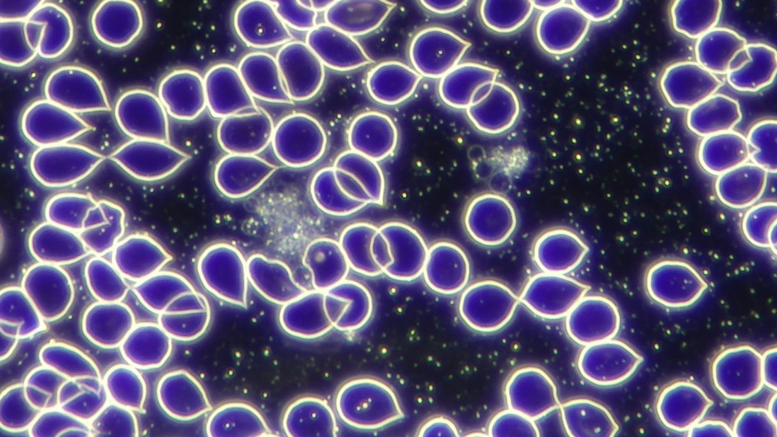
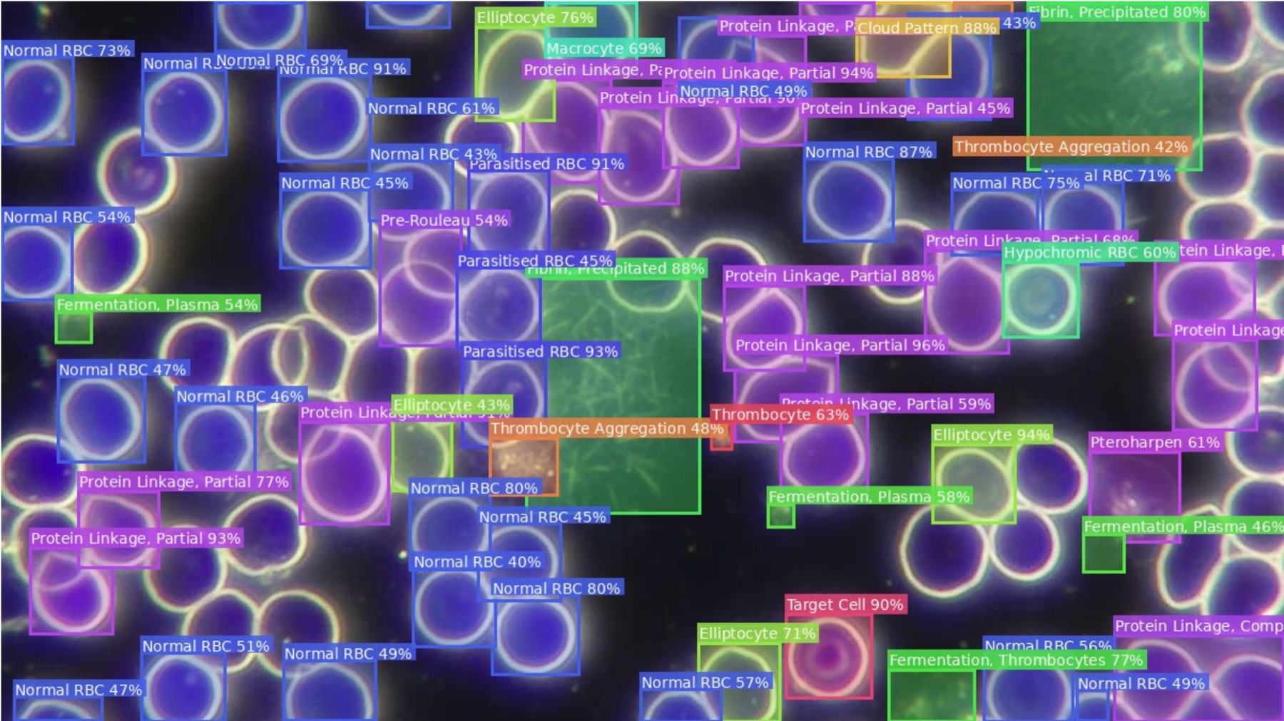
Raw capture → AI-annotated in minutes
Imagine this: your client sits across from you.
A single drop of blood goes under the microscope. Within minutes, your screen lights up — every cell automatically detected, labelled, and graded. Ghost cells, elliptocytes, cloud patterns, protein linkages — all identified with confidence scores. No guessing. No searching through reference books.
Before your client finishes their glass of water, you hand them a beautifully formatted report: ranked health priorities, a personalised action plan with dietary changes, supplement dosages, and lifestyle recommendations — plus the exact lab tests they should ask their doctor about.
From Blood Drop to Full Report
Follow along as a single darkfield microscopy image becomes a comprehensive practitioner report — in under 3–5 minutes.
1 · Capture or Upload
A finger prick, a slide, your darkfield microscope. Capture the fields live from your camera inside the app, or upload the JPG or PNG photos you already have — up to 20 per sample type. That's all you do.
2 · AI Detection, Your Review
A deep learning model trained on 1M+ cells scans every corner. Each cell gets a bounding box, a label, a confidence score, and a G1–G5 severity grade. Then you confirm: rename, fix a single cell, draw what it missed — nothing reaches the report without you.
3 · Health Priorities
Detections are cross-referenced against a knowledge base and scored into up to 30+ ranked health priorities — immune, nutritional, metabolic, and more.
4 · Action Plan
For each top priority: what to eat, which supplements at which dosages, which habits to adopt — with red-flag warnings for referral situations.
5 · Anisocytosis Analysis
Individual RBC diameters measured in micrometres, coefficient of variation calculated, and a colour-coded severity grade assigned automatically.
6 · Lab Test Bridge
Evidence-based lab test recommendations — CRP, ferritin, full blood count and more — prioritised by relevance, so clients can follow up with their GP.
What's in Every LiveBlood AI Report
AI-Annotated Blood Images
Every cell detected and labelled with bounding boxes, anomaly type, and confidence score — redrawn from your corrections, so the picture shows what you confirmed.
Health Priorities and Scoring
Up to 30+ health priorities ranked by severity, with colour-coded category tags and contributing anomaly breakdowns.
Medical Considerations
34+ medical perspectives scored and categorised — from cardiovascular to immune, metabolic to nutritional.
Action Plans
Structured dietary, supplement, and lifestyle recommendations for each top condition — with red flag warnings.
Diagnostic Investigations
Specific lab test recommendations for follow-up, prioritised by relevance to the findings.
Anisocytosis Analysis
Automatic RBC diameter measurement with coefficient of variation, severity grading, and visual colour-coded display.
LiveBlood AI vs. Manual Analysis
| Capability | LiveBlood AI | Manual Analysis |
|---|---|---|
| Time to generate report | ✓ Under 3–5 minutes | ✗ 30–90 minutes |
| Anomaly types detected | ✓ 50+ simultaneously | ✗ Limited by experience |
| Severity grading (G1–G5) | ✓ Automatic & consistent | ✗ Subjective estimation |
| Health implication scoring | ✓ AI-scored & ranked | ✗ Relies on memory |
| Action plan | ✓ Auto-generated per condition | ✗ Written from scratch |
| Annotated images with labels | ✓ Every cell labelled | ✗ Not standard |
| Anisocytosis measurement | ✓ Automated CV calculation | ✗ Manual estimation |
| Reproducibility | ✓ 100% consistent | ✗ Varies by practitioner |
| Bilingual (EN/ES) | ✓ Built-in | ✗ Manual translation |
The AI does the heavy lifting. You focus on your client.
Practitioners worldwide are already using LiveBlood AI to deliver faster, more consistent, and more comprehensive darkfield microscopy reports.
Start Free Trial